|
Taxonomically, the genus Staphylococcus is in the Bacterial family Staphylococcaceae, which includes three lesser known genera, Gamella, Macrococcus and Salinicoccus. The best-known of its nearby phylogenetic relatives are the members of the genus Bacillus in the family Bacillaceae, which is on the same level as the family Staphylococcaceae. The Listeriaceae are also a nearby family. Staphylococcus aureus forms a fairly large yellow colony on rich medium, S. epidermidis has a relatively small white colony. S. aureus is often hemolytic on blood agar; S. epidermidis is non hemolytic. Staphylococci are facultative anaerobes that grow by aerobic respiration or by fermentation that yields principally lactic acid. The bacteria are catalase-positive and oxidase-negative. S. aureus can grow at a temperature range of 15 to 45 degrees and at NaCl concentrations as high as 15 percent. Nearly all strains of S. aureus produce the enzyme coagulase: nearly all strains of S. epidermidis lack this enzyme. S. aureus should always be considered a potential pathogen; most strains of S. epidermidis are nonpathogenic and may even play a protective role in their host as normal flora. Staphylococcus epidermidis may be a pathogen in the hospital environment. Staphylococci are perfectly spherical cells about 1 micrometer in diameter. They grow in clusters because staphylococci divide in two planes. The configuration of the cocci helps to distinguish staphylococci from streptococci, which are slightly oblong cells that usually grow in chains (because they divide in one plane only). The catalase test is important in distinguishing streptococci (catalase-negative) from staphylococci, which are vigorous catalase-producers. The test is performed by adding 3% hydrogen peroxide to a colony on an agar plate or slant. Catalase-positive cultures produce O2 and bubble at once. The test should not be done on blood agar because blood itself contains catalase.
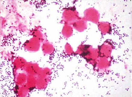
FIGURE 1. Gram stain of Staphylococcus aureus in pustular exudate Table 1. Important phenotypic
characteristics of Staphylococcus aureus
|